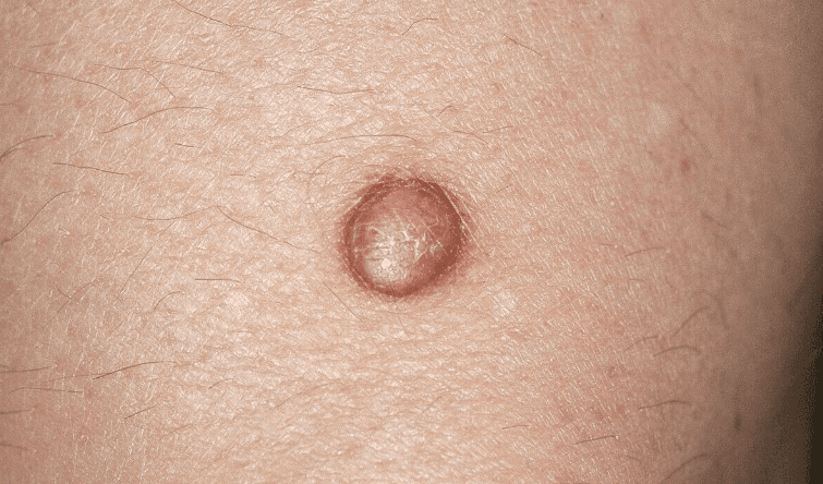
Dermatofibroma (also called histiocytoma) is a common benign skin growth arising from a localised proliferation of fibroblasts and histiocytes in the dermis. It develops most often on the lower limbs, occurs across all skin tones, and is more frequent in women — typically presenting in the 20–50 age range.
In many cases, dermatofibromas develop after minor skin trauma — an insect bite, needle prick, shaving cut, or thorn scratch. They are also more common in people with a weakened immune system, such as those living with HIV or autoimmune conditions.
Clinical Features
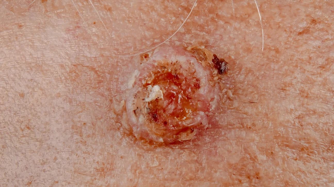
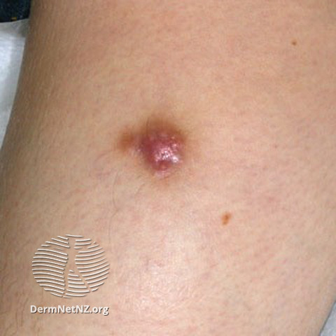
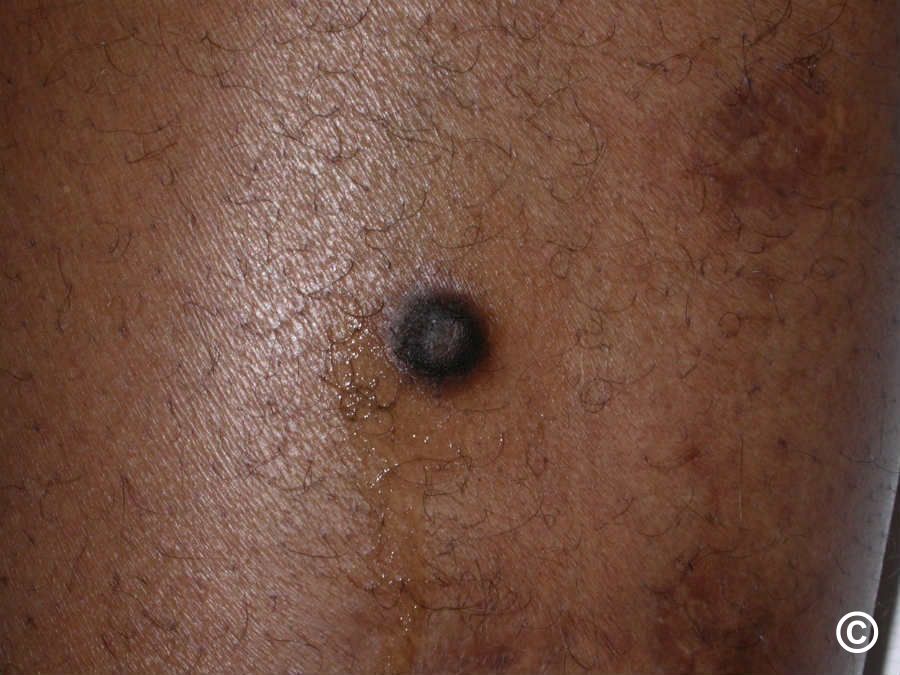
Dermatofibromas typically present as a firm, slightly raised or flat nodule with a smooth surface. Colour varies from pink or light brown on fair skin to dark brown or black on darker skin tones; a lighter area is sometimes visible at the centre. Size ranges from 0.5 to 1.5 cm, most commonly 7–10 mm.
Most lesions cause no symptoms. Some people, however, notice:
- A tightness or pulling sensation over the growth
- Pain or tenderness to touch
- Itching
- Increased local sensitivity
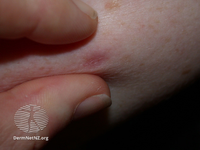
A reliable bedside finding is the dimple sign (pinch sign): gentle squeezing of the skin on both sides of the lesion causes the centre to dimple inward. This happens because the growth is tethered to the deeper layers of the skin.
Diagnosis
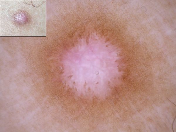
Dermatofibroma is usually diagnosed through clinical examination alone. Dermoscopy — a non-invasive magnified skin examination — helps a dermatologist assess the lesion's internal structure and distinguish it from other growths.
In atypical or uncertain cases, surgical excision with histological analysis (skin biopsy) provides a definitive diagnosis and removes the lesion at the same time.
Treatment
Because dermatofibromas are harmless, treatment is not always needed. Removal is considered when the growth causes persistent discomfort, when histological confirmation is required, or when the patient requests it for cosmetic reasons.
Available methods:
- Surgical excision — the most thorough approach; recurrence is uncommon but a visible scar usually remains
- Cryotherapy — liquid nitrogen freezing; lower scar risk, but higher chance of recurrence
- Electrosurgery — useful for superficial lesions
- Laser therapy — may reduce pigmentation but rarely removes the full depth of the growth
When to See a Dermatologist
Dermatofibroma can look similar to certain malignant skin tumours, particularly melanoma. Any skin growth that is new, enlarging, changing colour, or symptomatic warrants professional assessment. A dermatologist will use dermoscopy to evaluate the lesion and, if there is any doubt, arrange a skin biopsy to rule out malignancy definitively. Book an online dermatologist consultation if you have a skin growth you would like assessed.